Bone Cross Section / Compact Bone Decalcified Preparation Cross Section And Longitudinal Gsc Go Science Crazy. Find the perfect cross section bone stock photo. The central tubular region of the bone, called the diaphysis, flares outward near the end to form the metaphysis, which contains a largely cancellous, or spongy, interior. Derivative works of this file: Bone cross section view : 2 bone marrow makes more than 200 billion new blood cells.
Human bone, cross section diagram of femur showing osteon, veins, marrow. Start studying cross section of bone. Start studying bone cross section 2. Long bone diagram labeled colored 12 photos of the long bone diagram labeled colored , bone. Related posts of cross section of a long bone.
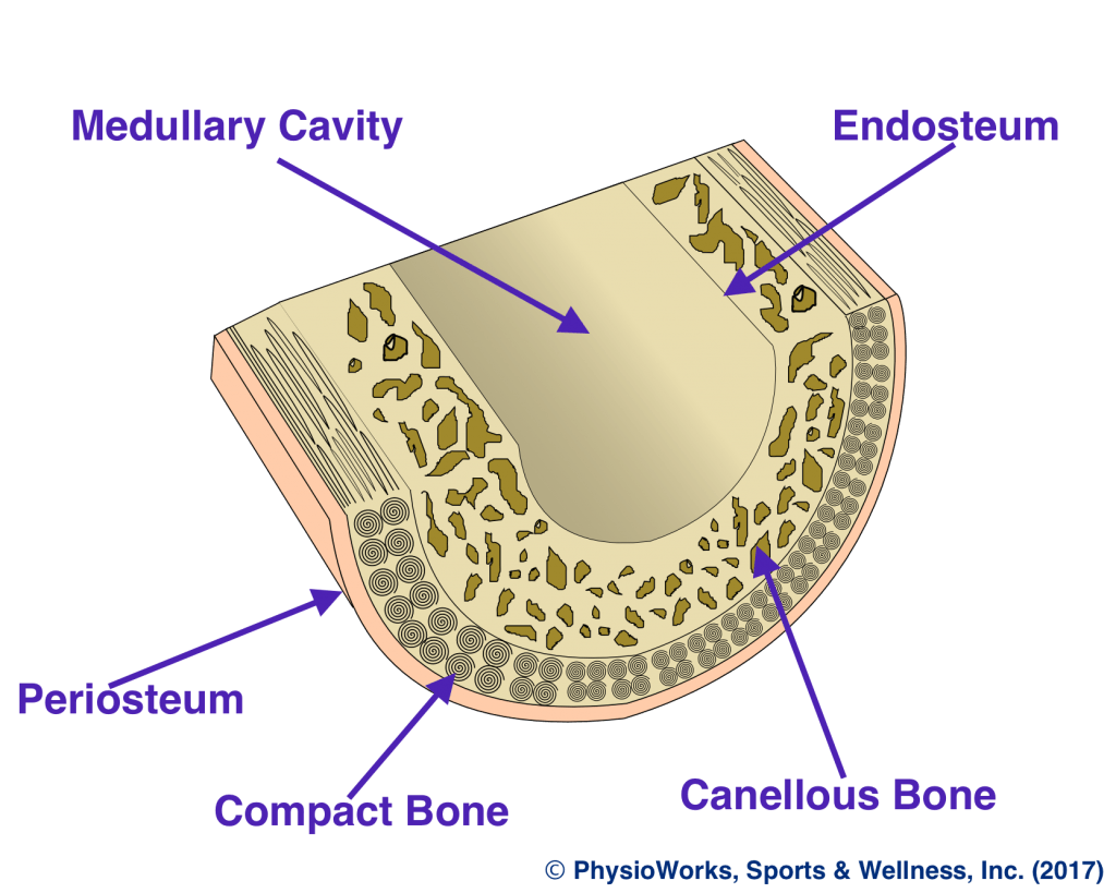
Human Skeletal System And Bone Cross Section For Classroom And Commercial Use from ecdn.teacherspayteachers.com Smartdraw includes 1000s of professional healthcare and anatomy chart templates that you can modify and make your own. Related posts of bone cross section labeled long bone diagram labeled colored. They are obtained by taking imaginary slices perpendicular to the main axis of organs, vessels, nerves, bones, soft tissue, or even the entire human body. Related posts of cross section of human bone diagram muscles and bones of the human body. At the end of the bone is the epiphysis, which in young people is separated from the. Derivative works of this file: Diagram with articular cartilage, marrow, spongy bone, medullary cavity, endosteum, diaphysis, and periosteum. The upper (biting) surfaces of the tooth are at top, with the lower sections (bottom) embedded in the gums and jaw bone (not shown).
A cross section of a human long bone.
Cross‐sectional area is derived from the integral of the bone mass profile across the narrow region. There is a printable worksheet available for download here so you can take the quiz with pen and paper. Internal structure of a human long bone, with a magnified cross section of the interior. It consists of two layers; Related posts of cross section of a long bone. They are obtained by taking imaginary slices perpendicular to the main axis of organs, vessels, nerves, bones, soft tissue, or even the entire human body. The upper (biting) surfaces of the tooth are at top, with the lower sections (bottom) embedded in the gums and jaw bone (not shown). If you are looking for the online quiz that this printable worksheet is based on, visit bone histology bone cross section. Smartdraw includes 1000s of professional healthcare and anatomy chart templates that you can modify and make your own. Two types of bone tissues in cross section of a long bone : Bone cross section view : This is an online quiz called bone cross section. No need to register, buy now!
An outer 'fibrous layer' containing mainly fibroblasts, and an inner 'cambium layer' containing progenitor cells. Derivative works of this file: Browse 53 bone marrow cross section stock photos and images available, or search for bone cross section or bone cells to find more great stock photos and pictures. As the names suggest compact bone looks compact and the spongy bone looks like sponges. Two types of bone tissues in cross section of a long bone :
Bone Stress Physioworks Sports And Wellness Inc from pwpull-bd87.kxcdn.com Compact bone is the outer layer and the spongy bone forms the inner layer. Internal structure of a human long bone, with a magnified cross section of the interior. A cross section of any bone will demonstrate these two types of bones. An outer 'fibrous layer' containing mainly fibroblasts, and an inner 'cambium layer' containing progenitor cells. The compact bone is made up of osteon. Two types of bone tissues in cross section of a long bone : Related posts of cross section of human bone diagram muscles and bones of the human body. There are two ways to study bone histology.
A cross section of a human long bone.
There is a printable worksheet available for download here so you can take the quiz with pen and paper. The compact bone is made up of osteon. Product is not alive nor is it edible. Huge collection, amazing choice, 100+ million high quality, affordable rf and rm images. Muscles and bones of the human body 12 photos of the muscles and bones of the human body anatomy bones of the human body quiz, major muscles and bones in the human body, muscles and bones in the human body, number of muscles and bones in the human body. Human bone, cross section diagram of femur showing osteon, veins, marrow. Start studying bone cross section 2. The central tubular region of the bone, called the diaphysis, flares outward near the end to form the metaphysis, which contains a largely cancellous, or spongy, interior. Explaned distal and proximal epiphysis. Vector illustration scheme of bone cross section. A cross section of a human long bone. Internal structure of a human long bone, with a magnified cross section of the interior. Find the perfect bone cross section stock photos and editorial news pictures from getty images.
Find the perfect cross section bone stock photo. No need to register, buy now! Learn vocabulary, terms, and more with flashcards, games, and other study tools. Explaned distal and proximal epiphysis. A property of the cross‐sectional area that represents the magnitude of the greatest bending rigidity of the section (cm 4).
Bone Cross Section Stock Photo Download Image Now Istock from media.istockphoto.com This is a short tutorial using blender 2.8 that shows how to create a bone cross section and using images to create the textures.hope you enjoy and please su. An outer 'fibrous layer' containing mainly fibroblasts, and an inner 'cambium layer' containing progenitor cells. Human bone, cross section diagram of femur showing osteon, veins, marrow. Browse 9,121 bone cross section stock photos and images available, or search for bone marrow or bone structure to find more great stock photos and pictures. If you are looking for the online quiz that this printable worksheet is based on, visit bone histology bone cross section. Items portrayed in this file depicts. Derivative works of this file: Select from premium bone cross section of the highest quality.
Diagram with articular cartilage, marrow, spongy bone, medullary cavity, endosteum, diaphysis, and periosteum.
Beautiful tooth cross section illustration, deep blue background and sparkling lights around. Select from premium bone cross section of the highest quality. The compact bone is made up of osteon. They are obtained by taking imaginary slices perpendicular to the main axis of organs, vessels, nerves, bones, soft tissue, or even the entire human body. They are obtained by taking imaginary slices perpendicular to the main axis of organs, vessels, nerves, bones, soft tissue, or even the entire human body. While it is not as hard as compact bone, spongy bone plays an important role of protecting the marrow where blood cells are produced. It consists of two layers; Product is not alive nor is it edible. English polish dutch ukrainian captions. 2 bone marrow makes more than 200 billion new blood cells. A property of the cross‐sectional area that represents the magnitude of the greatest bending rigidity of the section (cm 4). No need to register, buy now! A cross section of a human long bone.
Share :
Post a Comment
for "Bone Cross Section / Compact Bone Decalcified Preparation Cross Section And Longitudinal Gsc Go Science Crazy"


Post a Comment for "Bone Cross Section / Compact Bone Decalcified Preparation Cross Section And Longitudinal Gsc Go Science Crazy"