Back Muscle Chart - Low Back Muscles Anatomy Anatomy Drawing Diagram. The back's muscles start at the top of the back (named the cervical vertebrae) and go to the tailbone (also named the coccyx). Quadriceps (made of 4 muscles): This chart shows the outermost layer, called the superficial layer, of our major muscles. The muscles, bones, ligaments, and tendons in the back can all be injured and cause back pain. Back muscle diagram back muscles big back big back muscles big lats bodybuilding secrets major back muscles.
Most of the time, back muscle pain is diagnosed then treated with little more than a prescription of rest, painkillers and muscle relaxants. People with back pain people who experience headaches printing for use during doctor visits to communicate information about your symptoms quickly tracking your progress over time related tools: We hope this picture anatomy of back muscles diagram can help you study and research. Facebook twitter google+ linkedin stumbleupon tumblr pinterest reddit vkontakte share via email print. The superficial group, the deep group, and the intermediate group.
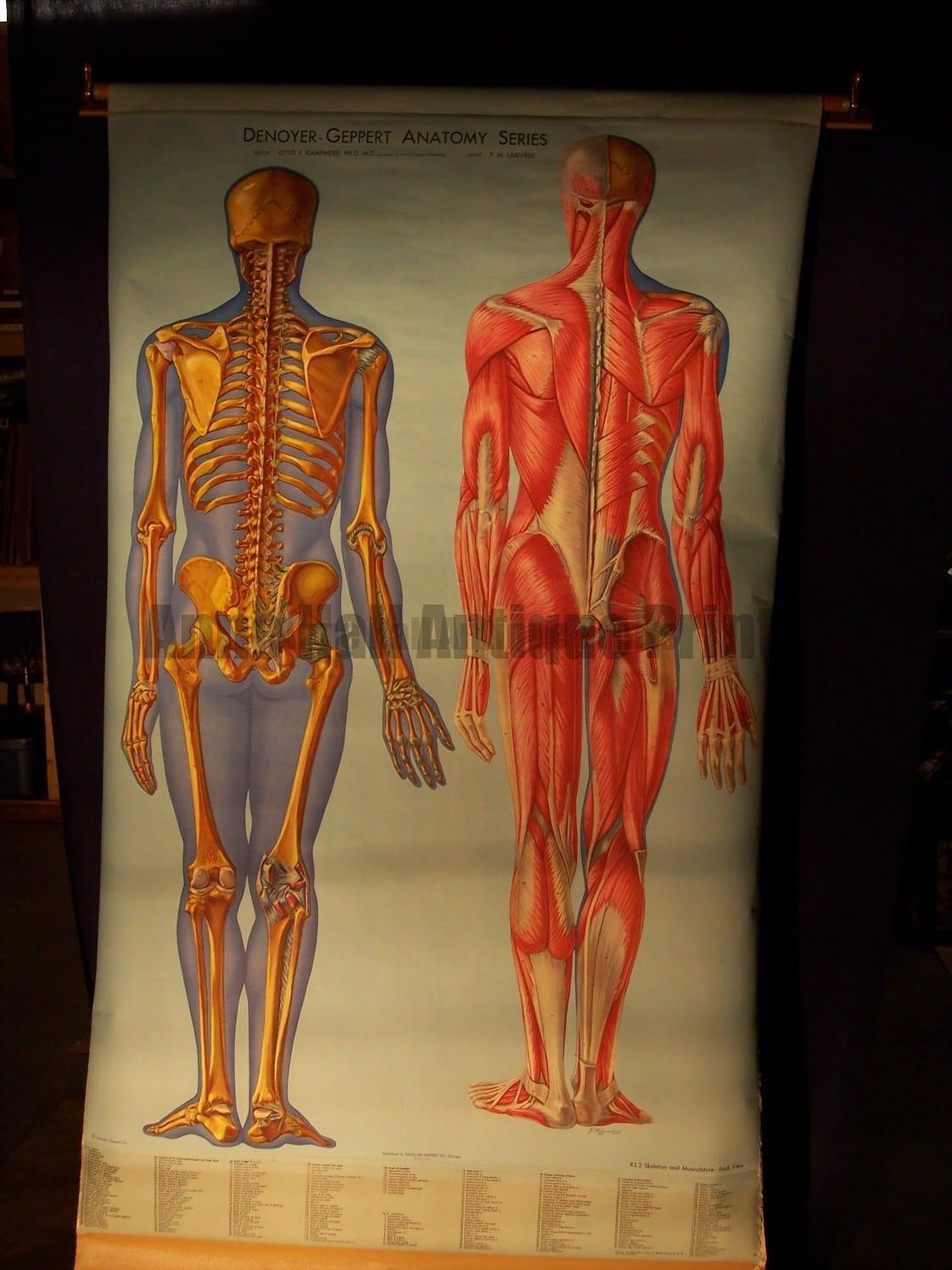
Anatomy Medical Vintage Teaching Charts Skeleton Muscles Brain Heart from annehallantiqueprints.com Another common cause of lower back and hip pain is disc injury. Muscle spasms (contraction or stiffening of the back muscles) muscles that feel tight; Human anatomy for muscle, reproductive, and skeleton. The muscles of the lower back help stabilize, rotate, flex, and extend the spinal column, which is a bony tower of 24 vertebrae that gives the body structure and houses the spinal cord. Chronic back pain map this tool recommended for: Vector illustration of human skeleton. Human muscle system, the muscles of the human body that work the skeletal system, that are under voluntary control, and that are concerned with movement, posture, and. This increases blood flow to the muscle normalizing it and bringing it back to a healthy state.
Intermediate back muscles and c.
We hope this picture anatomy of back muscles diagram can help you study and research. Muscle spasms (contraction or stiffening of the back muscles) muscles that feel tight; The muscles of the lower back help stabilize, rotate, flex, and extend the spinal column, which is a bony tower of 24 vertebrae that gives the body structure and houses the spinal cord. The most common type of back pain is muscle pain—also called muscle strain or soft tissue strain. The deltoid, teres major, teres minor, infraspinatus, supraspinatus (not shown) and subscapularis muscles (not shown) all extend from the scapula to the humerus and act on the shoulder joint. Muscles of the shoulder girdle posterior. This increases blood flow to the muscle normalizing it and bringing it back to a healthy state. Upper back muscles medical art library, labeled anatomy chart of neck and back muscles on white, diagram pictures superficial muscles of the back anatomy, interactive muscle chart back diagram. There are three different muscle groups found in the back: The back consists of the spine, spinal cord, muscles, ligaments, and nerves. For more anatomy content please follow us and visit our website: Some of these muscles are quite large and cover broad areas. Anatomynote.com found anatomy of back muscles diagram from plenty of anatomical pictures on the internet.
For the purposes of this feature, we're dividing the back into its four main regions: Certain back muscles extend to other areas, like the shoulders, upper arms, and thighs. The deltoid, teres major, teres minor, infraspinatus, supraspinatus (not shown) and subscapularis muscles (not shown) all extend from the scapula to the humerus and act on the shoulder joint. Other muscles are small and cover much less space. Pain log more pain mapping tools
Amazon Com Canvas On Demand Labeled Anatomy Chart Of Male Back Wall Decal Artwork Home Kitchen from m.media-amazon.com Another common cause of lower back and hip pain is disc injury. The intermediate layer contains the erector spinae muscles, whose many functions include the extension and lateral flexion of the spine, head and neck. Both the deltoid and the trapezius are firmly attached to the spine of the scapula. These structures work together to support the body, enable a range of movements, and send messages from the brain to. On this page, you'll learn about each of these muscles, their locations and functional anatomy. Others, like sumo deadlifts, have been shown in emg studies—and in the trenches—to focus more on other muscle groups than the back. This website uses cookies to improve your experience while you navigate through the website. However, the spinal erectors travel the length of the entire spine.
However, the spinal erectors travel the length of the entire spine.
People with back pain people who experience headaches printing for use during doctor visits to communicate information about your symptoms quickly tracking your progress over time related tools: Muscles found in the superficial group include rhomboid major, rhomboid minor, levator scapulae, trapezius, latissimus dorsi. Other muscles are small and cover much less space. Male reproductive organ simple diagram. Most of the time, back muscle pain is diagnosed then treated with little more than a prescription of rest, painkillers and muscle relaxants. The superficial group, the deep group, and the intermediate group. This is a diagram of the larger and more surface muscles of the low back. Facebook twitter google+ linkedin stumbleupon tumblr pinterest reddit vkontakte share via email print. To learn more about the anatomy of the spine, watch this video. The back isn't only one of the body's biggest and strongest body parts, it's also the most complicated in terms of being a series of interconnected muscle groups. Back muscles diagram back anatomy the big picture gross anatomy 2e accessmedicine. Human anatomy for muscle, reproductive, and skeleton. The muscles of the back can be arranged into 3 categories based on their location:
To download your free copy click the link. We hope this picture anatomy of back muscles diagram can help you study and research. Certain back muscles extend to other areas, like the shoulders, upper arms, and thighs. However, the spinal erectors travel the length of the entire spine. Some of these muscles are quite large and cover broad areas.
Muscle Diagram Of The Back Posterior Front Anterior from www.alpha-athlete.com Brings shoulders and arms back to body. The intermediate layer contains the erector spinae muscles, whose many functions include the extension and lateral flexion of the spine, head and neck. To download your free copy click the link. Anatomy chart courtesy of fcit the latissimus dorsi muscles (also known as the lats) are the largest muscles of the back. Function of the back muscles there are several individual muscles within the back anatomy, and it's important to take a quick look at all of The trapezius and latissimus dorsi muscles connect the upper limb to the vertebral column. We've created a free trigger point chart, which includes fybromyalgia treatment and reflexology information. We think this is the most useful anatomy picture that you need.
These structures work together to support the body, enable a range of movements, and send messages from the brain to.
Male reproductive organ simple diagram. Muscle spasms (contraction or stiffening of the back muscles) muscles that feel tight; Anatomy chart courtesy of fcit the latissimus dorsi muscles (also known as the lats) are the largest muscles of the back. Your clients will thank you for it! The back consists of the spine, spinal cord, muscles, ligaments, and nerves. Muscles are usually work in pairs because although they can contract and shorten (flex), they are pulled by an opposite (antagonist) muscle to straighten out (extend) again. Claim your free copy of the client back care guide today. They extend and rotate the head and neck. Back to tracking tools main page. We hope this picture anatomy of back muscles diagram can help you study and research. A strain can be an injury to a tendon attachment from muscle to bone. Leaning back to straight vertical and all points in between. Lower back muscle diagram anatomy does degenerative disc disease affect the lower back muscle?
Share :
Post a Comment
for "Back Muscle Chart - Low Back Muscles Anatomy Anatomy Drawing Diagram"


Post a Comment for "Back Muscle Chart - Low Back Muscles Anatomy Anatomy Drawing Diagram"